Back Muscles Anatomy : Labeled Anatomy Chart Of Male Back Muscles On Black Background Stock Photo Alamy / Muscles of the lumbar spine.
byAdmin•
0
Back Muscles Anatomy : Labeled Anatomy Chart Of Male Back Muscles On Black Background Stock Photo Alamy / Muscles of the lumbar spine.. These structures work together to support the body, enable a range of movements, and send messages from the brain to the. Anatomynote.com found anatomy of back muscles diagram from plenty of anatomical pictures on the internet. This curve, called lordosis, helps to: Leaning back to straight vertical and all points in between. The human spine is composed of 4 sections of vertebrae.
1 your spine in this region has a natural inward curve. For more anatomy content please follow us and visit our website: Both the deltoid and the trapezius are firmly attached to the spine of the scapula. The muscles of the lower back, including the erector spinae and quadratus lumborum muscles, contract to extend and laterally bend the vertebral column. The back consists of the spine, spinal cord, muscles, ligaments, and nerves.
Stijn Leidelmeyer Back Muscles Anatomy Study from cdnb.artstation.com 1) make midline incision along spines of vertebrae 2) extend from Ligaments hold the vertebrae in place, and tendons attach the muscles to the. The extrinsic back muscles are located in the back, but act to produce movements of the shoulder and assist respiration. Folge deiner leidenschaft bei ebay! They provide movements of the spine , stability to the trunk, as well as the coordination between the movements of the limbs and trunk. Together these muscles form a column, known as the erector spinae. Back muscles, functions and exercises: Muscle anatomy of forearm 12 photos of the muscle anatomy of forearm anatomy of forearm muscles pdf, human anatomy muscles of the forearm, muscle structure of forearm, muscle structure of the forearm, muscles of forearm anatomy ppt, human muscles, anatomy of forearm muscles pdf, human anatomy muscles.
Ligaments hold the vertebrae in place, and tendons attach the muscles to the.
These structures work together to support the body, enable a range of movements, and send messages from the brain to the. Muscle anatomy of forearm 12 photos of the muscle anatomy of forearm anatomy of forearm muscles pdf, human anatomy muscles of the forearm, muscle structure of forearm, muscle structure of the forearm, muscles of forearm anatomy ppt, human muscles, anatomy of forearm muscles pdf, human anatomy muscles. (2017, elsevier) should be consulted. 1) make midline incision along spines of vertebrae 2) extend from Anatomy chart courtesy of fcit the latissimus dorsi muscles (also known as the lats) are the largest muscles of the back. Understanding lower back anatomy is key to understanding the root of lower back and hip pain. Human musculature bodybuilding infographic muscular system vector human anatomy back muscle anatomy bicep male muscular anatomy human body anatomy female female anatomy muscle hamstrings muscle. The muscles of the back muscles make up a large part of the anatomy (structure) of the back. Folge deiner leidenschaft bei ebay! The spaces between the vertebrae are maintained by intervertebral discs that act like shock absorbers throughout the spinal column to cushion the bones as the body moves. The human spine is composed of 4 sections of vertebrae. They start at the top of the neck and go down to the tailbone. Anatomynote.com found anatomy of back muscles diagram from plenty of anatomical pictures on the internet.
Folge deiner leidenschaft bei ebay! Your lower back (lumbar spine) is the anatomic region between your lowest rib and the upper part of the buttock. Back muscle anatomy chart 12 photos of the back muscle anatomy chart back muscle anatomy chart, lower back muscle anatomy chart, human muscles, back muscle anatomy chart, lower back muscle anatomy chart. The back consists of the spine, spinal cord, muscles, ligaments, and nerves. The spaces between the vertebrae are maintained by intervertebral discs that act like shock absorbers throughout the spinal column to cushion the bones as the body moves.
Labeled Anatomy Chart Of Neck And Back Muscles On White Background Stock Photo Download Image Now Istock from media.istockphoto.com This curve, called lordosis, helps to: Ligaments hold the vertebrae in place, and tendons attach the muscles to the. Anatomy chart courtesy of fcit the latissimus dorsi muscles (also known as the lats) are the largest muscles of the back. These structures work together to support the body, enable a range of movements, and send messages from the brain to the. This blog post article is an overview of the muscles of the lumbar spine of the trunk. Anatomynote.com found anatomy of back muscles diagram from plenty of anatomical pictures on the internet. Über 7 millionen englischsprachige bücher. The lower back (where most back pain occurs) includes the five vertebrae in the lumbar region and supports much of the weight of the upper body.
The muscles of the back muscles make up a large part of the anatomy (structure) of the back.
Understanding lower back anatomy is key to understanding the root of lower back and hip pain. Both the deltoid and the trapezius are firmly attached to the spine of the scapula. These layers of back muscles help to mobilize and stabilize your trunk during your day to day activities. The lower back (where most back pain occurs) includes the five vertebrae in the lumbar region and supports much of the weight of the upper body. The human spine is composed of 4 sections of vertebrae. Leaning back to straight vertical and all points in between. All about the back muscles the back anatomy includes the latissimus dorsi, trapezius, erector spinae, rhomboid, and the teres major. We think this is the most useful anatomy picture that you need. The abdominal muscles also play a major role in the posture and stability to the body and compress the organs of the abdominal cavity during various activities such as breathing and defecation. What are the lower back muscles and their anatomy? These sections are cervical (neck), thoracic (upper and middle back), lumbar (lower back), and sacrum (tailbone). 1) make midline incision along spines of vertebrae 2) extend from Muscles of the lumbar spine.
We think this is the most useful anatomy picture that you need. These sections are cervical (neck), thoracic (upper and middle back), lumbar (lower back), and sacrum (tailbone). 1) make midline incision along spines of vertebrae 2) extend from Über 7 millionen englischsprachige bücher. (2017, elsevier) should be consulted.
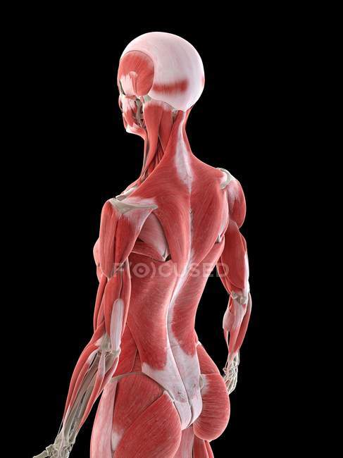
Female Anatomy Showing Back Muscles Computer Illustration Internal 3d Model Stock Photo 312138836 from st.focusedcollection.com It is a flat muscle that covers the largest area of the back and is the largest of its muscles, rather it is the largest muscle in the body. Back pain is common and might be caused by a problem with a muscle. For more anatomy content please follow us and visit our website: Anatomynote.com found anatomy of back muscles diagram from plenty of anatomical pictures on the internet. Understanding lower back anatomy is key to understanding the root of lower back and hip pain. Leaning back to straight vertical and all points in between. The trapezius and latissimus dorsi muscles connect the upper limb to the vertebral column. Back muscles, functions and exercises:
Muscle or ligament strains can occur from repeated use of the muscles, or from improperly or awkwardly lifting heavy objects.
For more anatomy content please follow us and visit our website: They start at the top of the neck and go down to the tailbone. Browse 3,565 back anatomy muscles stock photos and images available, or start a new search to explore more stock photos and images. The muscles, bones, ligaments, and tendons in the back can all be injured and cause back pain. Together these muscles form a column, known as the erector spinae. Browse 3,579 back muscle anatomy stock photos and images available, or search for pelvic bone or lymphatic system to find more great stock photos and pictures. This muscle extends from the bottom of the pelvis to the shoulders. These structures work together to support the body, enable a range of movements, and send messages from the brain to the. Back muscles the muscles of the back are a group of strong, paired muscles that lie on the posterior aspect of the trunk. Understanding lower back anatomy is key to understanding the root of lower back and hip pain. Back pain is common and might be caused by a problem with a muscle. We think this is the most useful anatomy picture that you need. The erector spinae is situated posterolaterally to spinal column, between the vertebral spinous processes and the costal angle of the ribs.